
New C3 Mutation, Linked to Severe aHUS, Found in Infant in Israel
Written by |
Khakimullin Aleksandr/Shutterstock


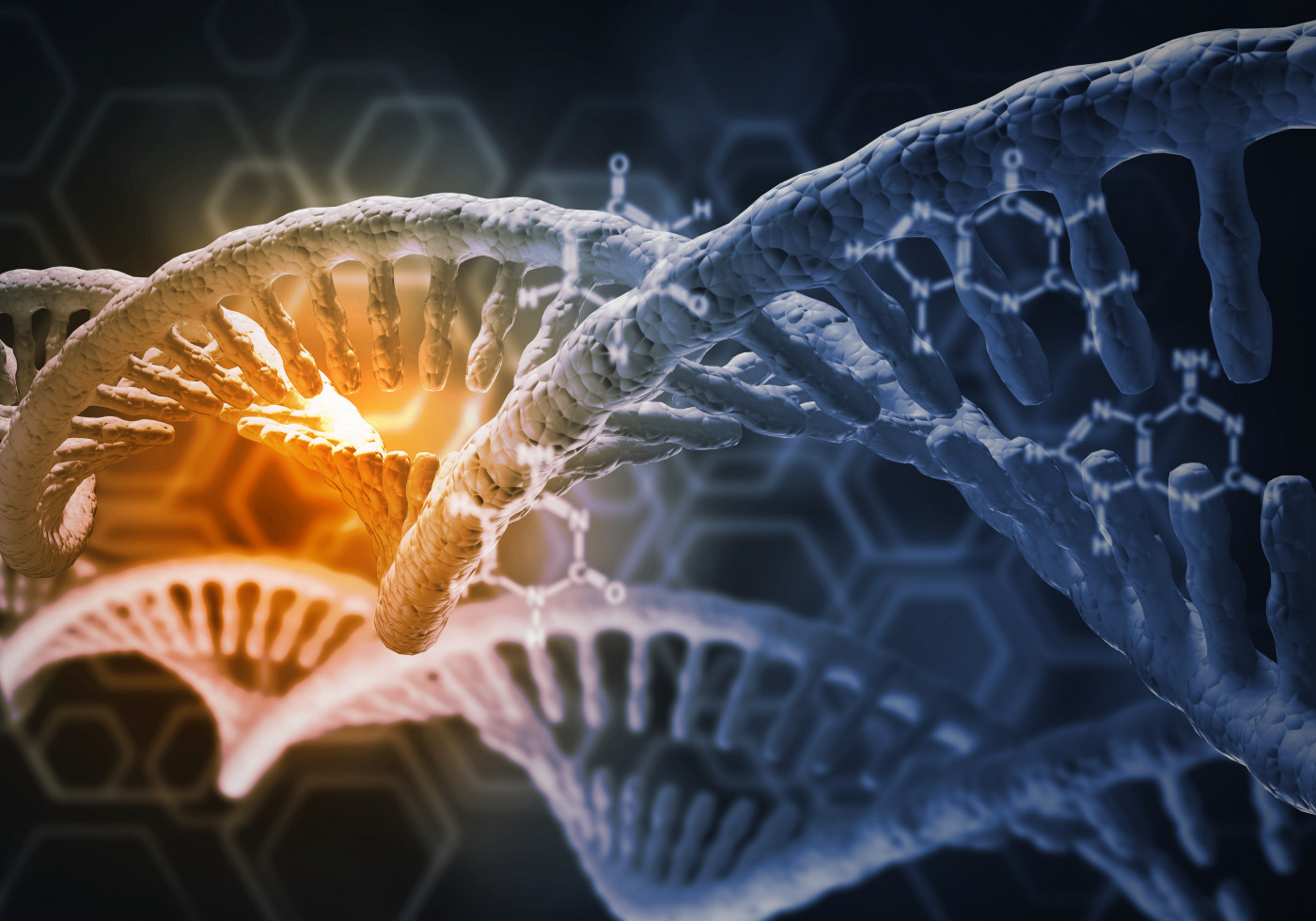
A newly identified mutation in the C3 gene that causes severe, life-threatening atypical hemolytic uremic syndrome (aHUS) was found in a baby boy in Israel, scientists report.
While C3 mutations leading to overactivation of complement protein C3 are associated with aHUS, they are usually found in only one of the gene’s two copies, known as heterozygous mutations.
The reported case is thought to be the first of a homozygous mutation, or a disease-causing mutation in both C3 gene copies, and one associated with severe neonatal presentation.
The case study, “A Novel Homozygous In-Frame Deletion in Complement Factor 3 Underlies Early-Onset Autosomal Recessive Atypical Hemolytic Uremic Syndrome – Case Report,” was published in the journal Frontiers in Immunology.
aHUS is characterized by the progressive destruction of red blood cells, low platelet counts, and subsequent tissue damage due to excessive activation of the alternative pathway of the complement system.
The complement system is a set of more than 30 blood proteins that form part of the body’s immune defenses. About two-thirds of aHUS patients carry aHUS-predisposing mutations in complement system genes.
C3 is the most abundant complement component protein, playing a crucial role in the activation of all complement pathways. Notably, aHUS secondary to abnormal C3 function is relatively rare, especially during childhood, accounting for 4.5–11.4% of aHUS cases.
C3-associated aHUS is mostly attributed to heterozygous gain-of-function variants in the C3 gene — meaning that they lead to an overly active C3 — and excessive complement system activation .
A team of researchers in Israel, along with colleagues in Hungary, identified a new homozygous C3 mutation in a baby boy that was associated with C3 overactivation and severe, early onset aHUS.
The boy — the fifth child of healthy, first-degree cousins of Arab-Muslim origin — was admitted to a local hospital in Israel when he was 3 days old due to extensive jaundice, anemia, low platelet counts, and impaired kidney function. Jaundice is a condition caused by the buildup of a yellow substance produced when red blood cells break down.
Following sudden kidney failure and respiratory insufficiency, he was transferred to the researchers’ hospital, where he needed mechanical ventilation and kidney replacement therapy.
Extensive testing eliminated other possible causes, and allowed the detection of persistently and extremely low blood C3 levels, with normal levels of the C4 complement protein — indicative of excessive activation of the alternative complement pathway. Of note, C3 only exerts its biological activity when converted into C3b; as such, low C3 levels typically reflect C3 consumption to produce its active form.
The overall symptoms and laboratory findings confirmed an aHUS diagnosis, and genetic testing looking for mutations in aHUS-associated genes identified a new C3 mutation (c.3322_3333del) present in both gene copies.
The mutation led to the deletion of four highly conserved amino acids — protein building blocks — in the resulting protein, thereby being considered disease causative.

The baby was also found to carry genetic changes in part of an aHUS genetic risk factor called MCPggaac, which is known to influence aHUS presentation in all carriers of C3 mutations.
Targeted sequencing of all healthy close relatives found none carrying the homozygous c.3322_3333del mutation, but the boy’s father, mother, sister, and one brother had the mutation in one C3 gene copy. His father and sister also carried the MCPggaac risk factor.
Notably, the boy’s mother showed intermediate C3 consumption and alternative pathway activation, suggesting that a heterozygous c.3322_3333del mutation may not cause full-blown disease, but be associated with milder abnormalities.
Further analyses of complement components and simulations of their interactions indicated that the boy’s mutated C3 protein lacked amino acids in its thioester-containing domain (TED), a region necessary for the interaction between its by-product C3b and FH, a suppressor of C3b’s subsequent activation.
These findings suggest that the c.3322_3333del mutation in both C3 gene copies results in a C3b protein that is resistant to inactivation by FH, thereby removing the breaks in C3’s activation cascade — and allowing for excessive alternative pathway activation and aHUS.
Following an aHUS diagnosis, the infant was treated with the complement suppressor Soliris (eculizumab), along with mechanical ventilation, daily hemodialysis, red blood cell transfusions, and preventive antibiotic therapy.
The boy showed rapid clinical and lab test improvements, and at 2 years old was in sustained disease remission under preventive treatment with Soliris. He had a history of two reversible aHUS relapses that followed a two-week delay in scheduled Soliris use.
“To the best of our knowledge, this is the first report of a homozygous deletion in C3 associated with severe neonatal presentation of aHUS, secondary to C3 over-activation,” the researchers wrote.
“Our findings enable prenatal diagnosis and early treatment initiation in newly identified subjects at risk, while reinforcing the significance of an intact C3-TED for normal function and regulation of the ACP [alternative complement pathway],” the team added.
Further research, they noted, is needed to clarify the exact mechanism whereby the newly identified C3 deletion may affect C3b molecular interactions with additional regulators of the alternative complement pathway.
Leave a comment
Fill in the required fields to post. Your email address will not be published.