Identical twins have same aHUS mutation but only 1 develops the disease
Non-genetic factors likely affect how aHUS is expressed, researchers say
Written by |

Two identical twins have the same disease-linked mutation in the complement factor B (CFB) gene, but only one developed atypical hemolytic uremic syndrome (aHUS), a recent study reports.
Despite only one having aHUS, the identical, or monozygotic, twins had similar levels of complement system activity — a part of the immune system that is abnormally active in aHUS — and similar concentrations of several complement activation molecules.
“The aHUS disease phenotype [manifestation] may vary considerably between carriers of the same mutation even in the same family and the affected individual’s genotype [genetic background] cannot solely explain [their] phenotype,” researchers wrote. “We therefore conclude that other, non-genetic factors, are likely to affect the expression of disease in these individuals.”
The report, “Factor B mutation in monozygotic twins discordant for atypical hemolytic uremic syndrome,” was published in the journal Kidney International Reports.
Trigger often needed for aHUS to develop
The complement system, comprising more than 30 blood proteins, is part of the body’s immune defenses. When activated in response to a threat, it triggers the activation of a powerful cascade of pro-inflammatory and blood clotting proteins.
aHUS is associated with mutations in genes that regulate the activity of the complement system. These mutations result in unwanted complement activation, leading to increased inflammation and formation of blood clots in small blood vessels, particularly in the kidneys.
However, just the presence of such mutations is typically not enough for the disease to develop. A trigger event, such as an infection, is often needed as well. This makes it difficult to define the risk of aHUS in families with members who carry disease-causing mutations.
In this study, a team of researchers in Iceland and Sweden investigated a large family in Iceland who has a history of aHUS with the goal of better understanding the importance of genetic mutations to the development of the disease.
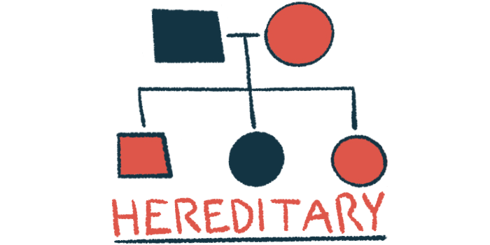
Three members of the family had aHUS: a father, identified as III-8, and his two sons (IV-9 and IV-10). All three had the same genetic mutation, D371G, in the CFB gene, with disease onset occurring at different ages and following varied courses.
These findings suggest that carrying the mutated variant, as such, does not fully account for disease expression but should be considered an important risk factor.
The father had one episode of severe kidney failure some weeks after having a flu-like illness that improved greatly after plasma exchange therapy. The older son had severe kidney failure requiring temporary dialysis, after which he recovered and has remained stable. The younger son was diagnosed with aHUS and kidney failure at a young age and has received three kidney transplants.
In addition to these three individuals, four other family members have the same CFB mutation, but did not develop aHUS. This mutation has not been found so far in other families, according to the researchers, who examined a large genetic database covering about a seventh of Iceland’s population. A genetic investigation of the extended family, covering 203 individuals over five generations, traced the mutation’s origin to an ancestral couple from the 19th century, or perhaps their parents.
One of the four unaffected mutation carriers in the family, III-7, is the monozygotic twin of III-8. Both have a similar body mass index, a measure of body fat. No other mutations or genetic modifications were found in the twin with aHUS. Two decades after III-8 first experienced symptoms of aHUS, his twin brother has not developed any signs or symptoms of the disease.
However, analyses of their blood serum — the liquid part of blood that does not contain blood cells or blood clotting proteins — revealed that both had elevated levels of several molecules resulting from complement system activation compared with the serum of healthy people.
A test of the serum’s ability to activate the complement system revealed a comparable result. Serum samples from both twins similarly induced the activation of the complement system in glomerular endothelial cells — specialized cells in the kidneys that play a key role in filtration.
Serum of twin with aHUS led to higher levels of red blood cell destruction
The researchers found that serum from the twin with aHUS led to significantly higher levels of red blood cell destruction (hemolysis) in an analysis using sheep red blood cells compared with serum from healthy individuals. Serum from the twin without aHUS also led to increased levels of hemolysis, but this was not statistically significant.
The research team hypothesized that these differences in the twins likely does not stem from a genetic difference, but rather from epigenetic variations, meaning reversible changes in how genes are used in the body.
Such epigenetic variations “may also be linked to environmental exposures such as pre- and postnatal events, infections, inflammations, vaccinations as well as lifestyle variations, including body mass index discordance, that may contribute to complement activation and differences between monozygotic twins,” the researchers wrote.
“These findings suggest that carrying the mutated variant, as such, does not fully account for disease expression but should be considered an important risk factor,” they added.


