Difficulties in Diagnosing aHUS When Family History Lacking Detailed in Case Report
Written by |


Difficulties in diagnosing atypical hemolytic uremic (aHUS) syndrome in the absence of family history are illustrated in the story of a girl in Sri Lanka who, after multiple diagnoses, was successfully treated for aHUS by long-term plasma exchange, a case study reported.
The study, “Atypical hemolytic uremic syndrome: a case report,” was published in the Journal of Medical Case Reports.
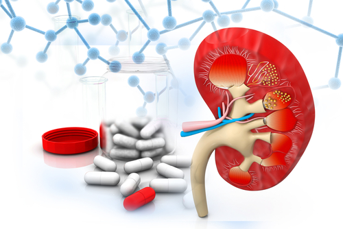
An aHUS hallmark is the formation of tiny blood clots (microthrombi) in small blood vessels that hamper blood flow to various organs, especially the kidneys.
The formation of clots in small vessels is called thrombotic microangiopathy (TMA). aHUS is considered to be one of three categories of TMA, each with similar symptoms but different causes.
aHUS can result from an overactive immune response triggered by an acute infection (30% to 50% of all cases are not genetic, the study notes), while Shiga toxin-associated hemolytic-uremic syndrome (Stx HUS) — the most common form of HUS — is caused by the infection of virulent bacteria. Thrombotic thrombocytopenia purpura (TTP) results from improper blood clot formation.
In the absence of family history, and its similarities to other conditions, a rare condition like aHUS can be overlooked. “Diagnosing aHUS is a challenge for a variety of reasons,” the authors said.
They detail a 15-year-old girl from Sri Lankan with no significant medical history, or family history of hypertension or diabetes, who was admitted to the hospital with a fever and skin rash.
She was diagnosed with the chickenpox virus (varicella), but later complained of severe abdominal pains, produced little urine, and had high blood pressure, a high temperature, and a fast heart rate.
An ultrasound confirmed a diagnosis of acute appendicitis along with acute kidney injury. She was treated with antibiotics and transferred to specialized care at the Teaching Hospital Kandy in Kandy, Sri Lanka, where her appendix was removed.
The day after surgery, she did not produce urine, her blood was acidic, and her creatine levels — a marker for kidney disease — rose. As a result, she started blood dialysis and her kidney was biopsied.
Three days later, she developed an infection, which was thought to be due to the surgery. However, no infectious agents were identified.
Further blood tests showed she had low hemoglobin and platelets, and blood film (smear) analysis revealed features of microangiopathy. With these test results and the presence of unexplained fever, TTP was suspected. She was admitted to the intensive care unit and started on a therapeutic plasma exchange.
Her condition was complicated by a lower respiratory infection and pulmonary hemorrhage, which triggered lung failure and required ventilation to help her breathe. Antibiotic treatment and plasma exchange continued until her urine output improved.
The kidney biopsy showed tissue damage in the small arteries. These findings together with high blood pressure, microangiopathies, pulmonary hemorrhage, and respiratory failure led to a final aHUS diagnosis.
The girl was given long-term plasma exchange, and she gradually responded to treatment. After 14 days in intensive care, she was discharged from the hospital and offered plasma exchange once a week for six months. She was gradually weaned from this treatment over 30 months.
After treatment stopped, her hemoglobin levels, platelet counts, creatinine levels, and blood film analysis were all normal.
“To our knowledge, there is only a handful of case reports on aHUS in Sri Lanka, and this is the only reported case with complete recovery following a lengthy course of plasma exchange in Sri Lanka,” the scientists wrote.
“Early diagnosis with prompt treatment will render a better outcome. The atypical hemolytic uremic syndrome needs to be considered in all patients with thrombotic microangiopathy,” they concluded.