Boosting Cell Stress Response May Be Therapeutic Approach in aHUS


Endothelial cells, or those that line blood vessels, are functionally abnormal in people with atypical hemolytic uremic syndrome (aHUS) associated with antibodies against complement factor H or CFH, according to a small study using patient-derived cells.
Notably, these intrinsic defects were associated with reduced activation of the p38 MAPK signaling pathway, which responds to cellular stress. When restored, this pathway rescued functional defects in lab-grown, patient-derived endothelial cells.
In addition, exposing these dysfunctional endothelial cells to patients’ blood containing anti-CFH autoantibodies aggravated their defects and promoted cell death, further supporting the idea that these antibodies represent a second “trigger” for developing full-blown aHUS in these patients.
These findings also point to p38 MAPK signaling as a potential therapeutic target for this subset of aHUS patients with anti-CFH autoantibodies, the researchers noted.
The study, “Patient-specific iPSC-derived endothelial cells reveal aberrant p38 MAPK signaling in atypical hemolytic uremic syndrome,” was published in the journal Stem Cell Reports.
aHUS is a rare disorder characterized by the destruction of red blood cells, low platelet levels, and the formation of blood clots in small blood vessels, which can lead to organ damage. It is caused by the abnormal activation of the complement system — a set of more than 30 blood proteins that form part of the body’s immune defenses.
Most patients with aHUS carry mutations in complement system genes that increase their predisposition to develop the disease. In some cases, aHUS has been associated with the production of antibodies that abnormally target proteins that prevent complement-mediated damage, such as CFH, ultimately leading to uncontrolled complement activation.
Anti-CFH autoantibodies-associated aHUS has been linked to genetic deletions in the CFHR1 and CFHR3 genes, which provide instructions to produce CFH-related proteins.
Endothelium damage due to excessive complement activation is thought to be a key event in aHUS, but “the role of endothelial cell alterations in aHUS has not been well characterized and the underlying mechanisms remain unclear,” the researchers wrote.
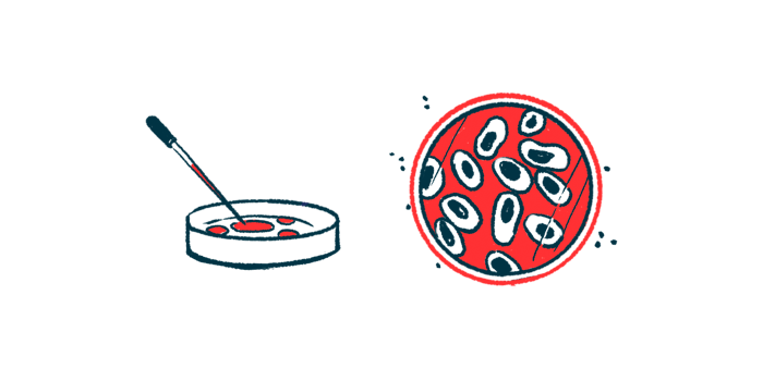
To address this, a team of researchers in China analyzed endothelial cells derived from three boys with anti-CFH autoantibodies-associated aHUS, of whom two carried genetic deletions in the CFHR1 and CFHR3 genes.
These cells were generated from patient-derived induced pluripotent stem cells (iPSCs) and grown in the lab. iPSCs are generated from fully matured cells that are reprogrammed back to a stem cell-like state, where they can give rise to almost every type of human cell.
As such, when derived directly from patients, these cells can be used as cellular models that mimic the disease’s genetic and clinical diversity. Endothelial cells also were derived from iPSCs of two boys who did not have the disease to be used as controls.
The results showed that endothelial cells from all three patients had functional defects, including a reduced ability to migrate, form tubes, and grow, relative to healthy cells. However, no differences in cell death were found between patient-derived and control cells.
These findings suggest “that an intrinsic defect in ECs [endothelial cells] occurs in anti-CFH autoantibody-associated aHUS,” regardless of CFHR1/CFHR3 mutations, the researchers wrote.
Endothelial cells from both patients and controls were exposed to aHUS patients’ blood, which contained anti-CFH autoantibodies. Following that, all of the cells showed impaired migration and tube formation, as well as increased cell death, the researchers said.
This observation supported the damaging effect of circulating anti-CFH autoantibodies and suggested that these abnormal antibodies worsen these patients’ intrinsic endothelial cell defects.

The team then investigated the potential causes of this inherited endothelial dysfunction, or impairment, by analyzing changes in gene activity. They found that 1,401 genes were differentially activated in patient-derived cells, compared with controls: 659 genes had higher activity, while 742 had lower activity.
Notably, most of the suppressed genes in patient-derived cells were associated with blood vessel-related processes.
Further analysis revealed that endothelial cells from all three patients had reduced p38 MAPK signaling, a pathway typically activated in response to cellular stress and essential for endothelial cell function.
Importantly, boosting p38 MAPK signaling, either pharmacologically or by genetic manipulation, in endothelial cells derived from all three patients rescued their defects, “indicating a common signaling pathway in anti-CFH autoantibody-associated aHUS and thus providing a new target for treatment of the disease,” the team wrote.
Anti-CFH autoantibody-associated aHUS patients “with or without CHFR1/3 deletion showed endothelial dysfunction and decreased p38 MAPK signaling, which might indicate that CHFR1/3 deletion was not associated with primary endothelial dysfunction and may provide a new explanation of the hereditary susceptibility of the disease onset,” they wrote.
This is consistent with the “two-hit” hypothesis of aHUS development, which considers that a genetic predisposition — the first “hit” — may not be sufficient to cause the full-blown condition, thereby requiring a second “hit,” which in this case may be the presence of anti-CFH autoantibodies.
Given that only two patients had CFHR1/CFHR3 mutations, the genetic predisposition that promotes the initial endothelial defects in these three patients may remain unknown.
The data also validated iPSC-derived endothelial cells as a reliable model to recapitulate disease-associated features, “thus providing a unique platform for gaining mechanistic insights into EC injury in aHUS,” the researchers wrote.