Blood disorder was likely trigger event for aHUS in woman, 38: Report
First reported case successfully treated with steroids, plasma exchange
Written by |


A group of blood disorders called hypereosinophilia syndrome, known as HES, was suspected to be the trigger event that led to atypical hemolytic uremic syndrome (aHUS) in a woman in her late 30s, according to a new case study.
HES is caused by an overload of eosinophils, a type of white blood cells, and can lead to organ damage.
“We reported the first case of HES followed by aHUS in a patient with underlying genetic complement factor I (CFI) deficiency,” the researchers wrote.
“The patient was successfully treated with high-dose steroids and extended duration of plasmapheresis,” or plasma exchange, the team added.
The case was reported in a study, “Complement Factor I Gene Variant in an Atypical Hemolytic Uremic Syndrome Triggered by Hypereosinophilia Syndrome,” published in the journal Nephron.

Trigger event usually needed in cases of aHUS
aHUS is caused by abnormal activity of the complement cascade, a part of the immune system, that leads to inflammation and blood clotting in small blood vessels, or thrombotic microangiopathy. This can damage organs, especially the kidneys.
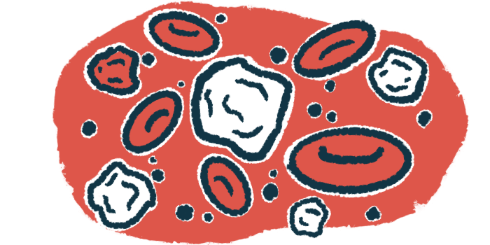
Other disease hallmarks include low platelet counts, called thrombocytopenia, and destruction of red blood cells — known as hemolytic anemia.
Most people with aHUS have mutations in genes that regulate the function of the complement cascade; however, a trigger event usually is also necessary. Frequently described triggers include autoimmune diseases, infections, pregnancy, and medications.
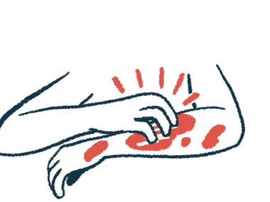
In this report, researchers in Thailand describe the case of a 38-year-old woman who sought treatment at a clinic for prolonged fever and rashes.
Three weeks prior, she had started having high-grade fever and hives (urticaria) on the chest, back, hands, and feet. She tried treatment with over the counter antihistamines and steroid creams, among other remedies, but with no improvement.
An initial physical examination revealed the woman had a body temperature of 38.5 C (101.3 F), normal blood pressure and heart rate, and angioedema, or swelling under the skin.
Lab tests showed the patient had mild anemia — with low hemoglobin, the protein that carries oxygen in red blood cells — and higher than normal levels of eosinophils. She had normal levels of platelets and of the C3 and C4 proteins of the complement system, as well as normal kidney and liver functions.
On day 11, the patient had convulsions, but MRI did not reveal brain damage. Further examination revealed high levels of eosinophils in the cerebrospinal fluid, which is the liquid that surrounds the brain and spinal cord, and in the bone marrow. The findings overall led to a diagnosis of HES with neurological involvement.

Researchers can’t rule out that disease co-occurrence was coincidence
The woman was started on a high dose of dexamethasone, a corticosteroid, but her consciousness and kidney function worsened. She required intubation and temporary hemodialysis — where a specialized machine performs the normal kidney filtering functions.
Further lab analysis, conducted on day 12, revealed a worsening of her anemia and thrombocytopenia, and an increase in the blood levels of lactate dehydrogenase — a measure of tissue damage. The analysis also found schistocytes, or fragments of red blood cells, and eosinophil degranulation. This release of mediators stored in eosinophil granules is a key event in chronic eosinophilic diseases.
The patient’s levels of the complement system proteins C3, C4 and CH50 were close to the minimum normal level. A kidney biopsy revealed thrombotic microangiopathy.
After not responding to intravenous or into-the-vein immunoglobulin therapy, she was diagnosed with aHUS.
Genetic analysis revealed a mutation in the CFI gene, associated with a deficiency in complement factor I, “which probably made her susceptible to developing aHUS,” the team wrote.
The variant is not found in population databases and is categorized by the American College of Medical Genetics as pathogenic moderate.
The woman was treated with plasma exchange, or plasmapheresis, and dialysis on alternate days. After 15 sessions of plasma exchange, her platelet levels gradually increased. Urine output also increased, enabling her to stop hemodialysis.
She regained consciousness, and the tube used to assist breathing was removed.
Because eosinophilia can lead to thrombosis from various mechanisms, we proposed that hypereosinophilia could be considered a trigger of aHUS in underlying complement-dysregulated patients.
At a 2.5-year follow-up, her kidney function was stable, and she had no recurrence of HES nor of thrombotic microangiopathy.
“Because eosinophilia can lead to thrombosis from various mechanisms, we proposed that hypereosinophilia could be considered a trigger of aHUS in underlying complement-dysregulated patients,” the researchers concluded.
“However,” they added, “the possibility that these two diseases coincidentally occurred cannot be excluded.”