aHUS Linked to Similar Disorder via Complement System and Protein Deficiency, Study Says
Written by |

Two diseases often difficult for doctors to distinguish between — atypical hemolytic uremic syndrome (aHUS) and thrombotic thrombocytopenic purpura (TTP) — are both marked by complement system activation and severe deficiency of the ADAMTS13 enzyme, researchers report.
These disorders are known to share symptoms, but until now were largely thought to be have distinct biological mechanisms.
The study, “Synergistic effects of ADAMTS13 deficiency and complement activation in pathogenesis of thrombotic microangiopathy,” was published in the journal Blood.
aHUS and TTP are two types of thrombotic microangiopathy, characterized by blockages in small blood vessels (thrombotic microangiopathy) that result in organ injury. While TTP is caused by autoantibodies against ADAMTS13 or inherited mutations in the ADAMTS13 gene, aHUS results from uncontrolled activation of the complement system, a set of over 20 blood proteins that form part of the body’s immune defenses.
But complement activation has also been shown in TTP, suggesting a link between the disorders. TTP patients with refractory disease and severe ADAMTS13 deficiency respond to Soliris (eculizumab, by Alexion Pharmaceuticals), a treatment that targets the complement C5 protein.
Researchers at the University of Alabama at Birmingham and the University of Pennsylvania intended to better assess if ADAMTS13 deficiency and complement activation both contribute to disorders such as aHUS and TTP.
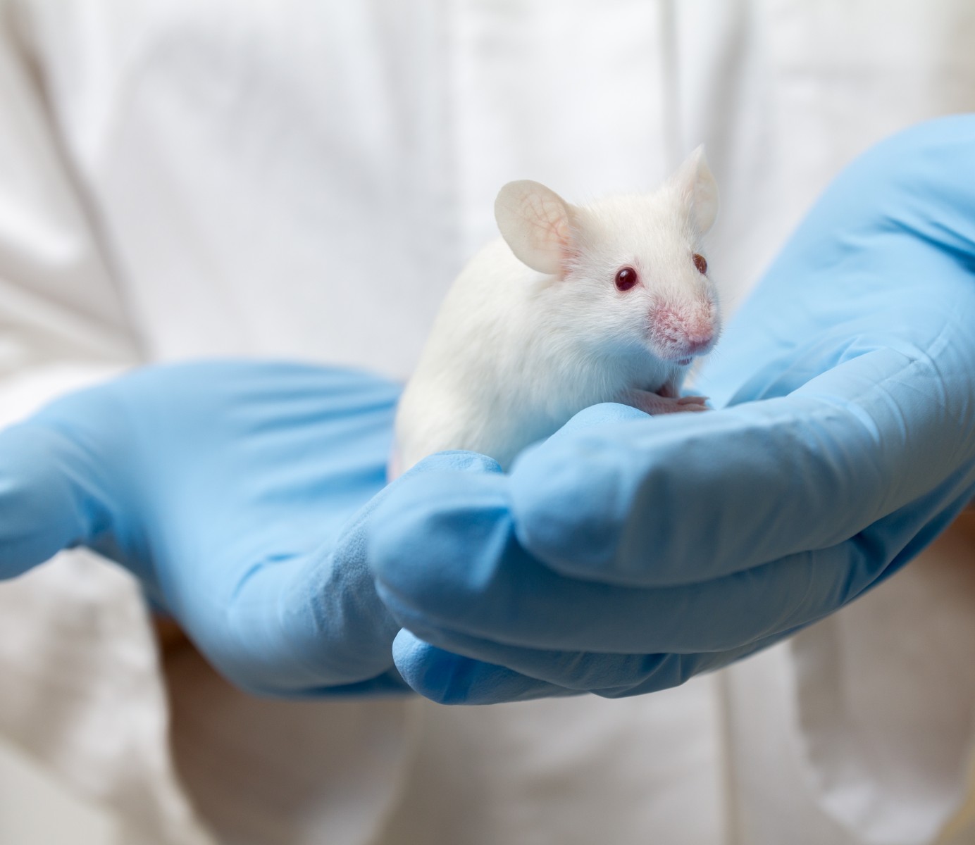
For this purpose, they generated mice with a combined mutation in Adamts13 and cfh, a gene that codes for the CFH complement protein and whose variants have been linked with aHUS.
Results revealed that mice with no ADAMTS13 protein or with a mutation in one of the copies (heterozygous) of cfh — resulting in an amino acid substitution of tryptophan by arginine — remained asymptomatic, despite having microvascular blood clots in various tissues.
In contrast, the animals lacking both the ADAMTS13 protein and with a heterozygous cfh mutation showed a higher mortality rate and more severe organ damage.
These mice also showed low levels of platelets (reflecting the formation of blood clots), increased amounts of the LDH enzyme (an indicator of overall organ damage), and higher levels of blood urea nitrogen and of the kidney function marker creatinine. The analysis further showed low plasma levels of the haptoglobin protein, an indicator of hemolysis (red blood cell destruction).
Mice with mutated cfh in both gene copies (homozygous) revealed severe thrombotic microangiopathy irrespective of having alterations in Adamts13. They also had greater complement activation and markedly higher plasma levels of the Willebrand factor, which plays a crucial role in TTP disease processes.
The animals with combined mutations showed similar changes, but greater severity of organ damage and increased mortality than those with changes in cfh alone.
All three affected mouse strains had widespread platelet-rich blood clots in small arteries and capillaries across tissues of major organ, including the brain, heart, lungs, kidney, liver, and intestine.
“Together, our results support a synergistic effect of severe ADAMTS13 deficiency and complement activation in pathogenesis of thrombotic microangiopathy in mice,” the team wrote.
“The results may help design a potentially novel strategy to treat the refractory thrombotic microangiopathy,” X. Long Zheng, MD, PhD, the study’s senior author, said in a press release.


