
Rare genetic variant linked to woman’s pregnancy-related aHUS
Case report says variant could be new genetic factor that may induce disease
Written by |




A rare genetic variant in the F2 gene may have contributed to the development of atypical hemolytic uremic syndrome (aHUS) and associated kidney damage in a Chinese woman who had just given birth, a study showed.
Although genetic testing revealed no known aHUS-causing mutations, the newly identified F2 variant, which was shown to increase levels of complement-related proteins, may represent “a new genetic factor that may induce aHUS,” the researchers wrote. The complement cascade, a part of the immune system, becomes overactive in aHUS.
Further research is needed to confirm this genetic link.
The study is titled “Pregnancy-associated aHUS with a non-complement variant: a case report. Can a coagulation gene variant dictate prognosis?” It was published in BMC Nephrology.

Pregnancy a common aHUS trigger
In aHUS, the complement system becomes overactive and causes blood clots to form in small blood vessels, including those in the kidneys. Although certain mutations in genes encoding complement proteins can predispose someone to aHUS, a trigger is typically needed for symptoms to develop. Pregnancy is a common trigger.
In this report, the researchers described the case of a 31-year-old woman whose pregnancy-associated aHUS may have been linked to a rare variant in the F2 gene, which encodes prothrombin, a protein in the blood that helps it to clot properly.
The woman had a history of high blood pressure during pregnancy and HELLP, a syndrome that involves destruction of red blood cells, low clot-forming platelets, and damage to the liver. Her kidney function was normal. During her previous pregnancy, she had experienced bleeding after giving birth.
Four minutes after her placenta was delivered at 38 weeks of gestation, her blood pressure dropped sharply and her heart rate increased. Her oxygen levels also dropped, while blood tests showed signs of problems in the blood’s ability to clot.
After excluding other potential causes, the doctors diagnosed her with an amniotic fluid embolism complicated by intravascular coagulation. The former occurs when the amniotic fluid, the fluid that surrounds the baby in the womb, triggers a strong immune response after entering the mother’s circulation. Intravascular coagulation is marked by widespread blood clotting that uses up platelets and clotting proteins, increasing the risk of bleeding.
Doctors stabilized the woman with a breathing tube, mechanical ventilation, intravenous (into-the-vein) fluids, medications to lower her blood pressure, and transfusions of red blood cells and fibrinogen, a protein needed for blood to clot. She was then transferred to the intensive care unit.

Woman’s condition worsens
The following day, she stopped producing urine, showed further signs of kidney problems, and developed seizures. Her blood levels of creatinine, a waste product used to assess kidney function, more than tripled. She also developed anemia (low red blood cells) and thrombocytopenia (low platelets).
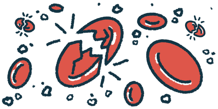
Blood tests revealed schistocytes, which are fragmented red blood cells caused by damage within small blood vessels. In addition, activity and levels of complement proteins were lower than normal, suggesting abnormal complement system activation.
Two days after giving birth, the woman developed a fever and cough and received antibiotics and antifungal medication for suspected infections. About a week later, she continued to show signs of kidney problems, as well as anemia and thrombocytopenia.
Blood transfusions and dialysis, a procedure that filters waste products from the blood when the kidneys cannot function properly, did not improve her condition.
A kidney biopsy — a small piece of kidney tissue collected for examination under a microscope — showed dead tissue in the outer part of the kidneys and their tube-like filtering units, as well as small blood clots.
“Combined with [tissue analysis] and laboratory findings, aHUS was clinically suspected,” the researchers wrote.
F2 variant increased activity of certain complement-coding genes
The woman began plasma exchange three times weekly to replace plasma, the liquid part of the blood that contains complement and complement-regulatory proteins, with fresh plasma. She also received alprostadil to lower blood pressure and rivaroxaban, a blood thinner.
After 10 plasma exchange sessions, destruction of red blood cells decreased. Platelets and complement proteins returned to normal. Her kidney function improved enough to stop dialysis.
She was discharged and continued long-term rivaroxaban because doctors suspected an increased tendency to form blood clots. Over six years of follow-up, her kidney function remained stable.
The evidence of complement activation despite the lack of genetic variants had prompted us to look beyond genetics, shifting the focus to the role of the coagulation [clotting] system in activating the complement system.
Genetic testing found no mutations in known complement regulatory genes but identified a variant called c.593G>A in one copy of the F2 gene.
“We hypothesized that the variant in F2 gene contributed to complement activation which caused aHUS,” the researchers wrote.
Experiments using lab-grown human cells that line blood vessels showed that the F2 variant increased the activity of certain complement-coding genes. The team hypothesized that the variant may make thrombin more active, thereby increasing both blood clotting and activation of the complement system.
“The evidence of complement activation despite the lack of genetic variants had prompted us to look beyond genetics … shifting the focus to the role of the coagulation [clotting] system in activating the complement system,” the researchers wrote.



Leave a comment
Fill in the required fields to post. Your email address will not be published.