 Discussion

Newly Diagnosed: Taking the First Step on Your Journey
Becoming educated is a good place to start on your journey with atypical hemolytic uremic syndrome (aHUS). Whether you are a patient or a caregiver, knowing as much as possible about the disease will help you be a more active participant in your or your loved one’s healthcare. Learn more below about aHUS, its causes, symptoms, and how it’s diagnosed.
Overview
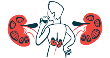
Atypical hemolytic uremic syndrome (aHUS) is a rare disease in which abnormal activity of the complement cascade, a part of the immune system, causes blood clots to form in small blood vessels. These clots cause damage to internal organs, especially the kidneys. aHUS is also a type of thrombotic microangiopathy, a group of diseases characterized by the formation of blood clots in the body’s small blood vessels, leading to organ damage.
Causes
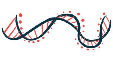
aHUS is caused by the abnormal activity of the complement cascade, which leads to increased inflammation and blood clotting in small blood vessels, particularly those in the kidneys. Over time, these small blood vessels can become clogged, leading to kidney damage and, eventually, kidney failure. Most people with aHUS have inherited mutations in the genes that regulate the function of the complement cascade.
Symptoms
The signs and symptoms of aHUS usually appear suddenly, and often follow a trigger event, such as an infection. There are three hallmark symptoms that define aHUS: hemolytic anemia, thrombocytopenia, and kidney failure. Symptoms can appear at any age, though it is slightly more common for them to first appear in childhood rather than later on in life. Adult-onset aHUS is more frequent in females than males, whereas childhood-onset disease affects both sexes equally.
Diagnosis
aHUS also is a type of thrombotic microangiopathy (TMA), which comprises a group of disorders characterized by the formation of blood clots in the body’s small blood vessels. A major challenge in diagnosing aHUS is differentiating it from other types of TMA. A main feature used to diagnose TMAs is evidence of hemolytic anemia in laboratory testing, most notably the presence of schistocytes (pieces of destroyed red blood cells) on a peripheral blood smear.